Comparison of a healthy dog with a high degree tvd-sick dog as an illustration of values:

Finding sheet of the heart examination of the Collegium Cardiologicum e.V. from Bysterstorff's Avalon Artus (left)
and Bysterstorff's Beam me up Schröder (right)
Based on the findings from Artus (free, negativ) and Schröder (high degree of tricuspid valve dysplasia) we would like to illustrate with numbers the impacts of this disease to the heart, especially to the right half of the heart (in particular to the right atrium):
The numbers marked red are those that are relevant for us.
Explanation:
LVDd – Left ventricle diameter in diastole
LAs – Diameter of the left atrium
RAs – Diameter of the right atrium
RVDd – Left ventricle diameter in diastole
What is it that you can see on those two finding sheets?
If you look at the numbers from the left half of the heart (LVDd and LAs) you can see that Artus diameter in the left ventricle is LVDd = 41.1mm and the diameter of the left atrium is LAs = 39.9mm. If you look at Schröders numbers you see that those are lower than those of Artus (LVDd = 32.4 and LAs = 31.6mm) which means that his left half of the heart is compared to a healthy heart smaller or rather is pushed back by the right half of the heart.
When you look at the right half of the heart (RVDd and RAs) you can see that Artus diameter of the right ventricle is RVDd = 21.1mm and the diameter of the right atrium is RAs = 24.6mm. You will recognize immediately that Artus right half of the heart is clearly smaller than his left side of the heart – this is how it is supposed to be! However Schröders right ventricle is RVDd = 33.9mm and his right atrium is RAs = 33.9mm which means it is very enlarged. You can see two things: On the on hand Schröders left ventricle and his right atrium are compared to Artus healthy heart heavily enlarged. On the other hand the ratio of Schröders right and left halves of the heart in comparison are extremely postponed. His left half of the heart is a small bag on the right half of the heart not as it should be the other way round (see Artus).
With those two finding sheets you can see very clearly what tricuspid valve dysplasia characterizes and how it affects the dog. The big to very big enlargement of the right half of the heart is typical of this kind of heart disease.
Comparison of a healthy dog with a high degree tvd-sick dog via ultrasound:
Using a cardiac ultrasound video from Artus (healthy, without clinical evidence) and Schröder (profoundly TVD) we like to demonstrate the effects of this disease visually.
First, two videos about the differences in size of the hearts via simple ultrasound, second two videos about the blood backflow in case of Schröder in comparison to the healthy Artus, using color Doppler ultrasound.
Illustration of size differences:
To begin with, we like to demonstrate the size differences of both hearts, the heart ventricles respectively. Here, we show one ultrasound-video (1-minute repeat mode) per dog, which were kindly provided by the cardiologist Dr. Kattinger. We also highlight the important parts on a screenshot for a better understanding.
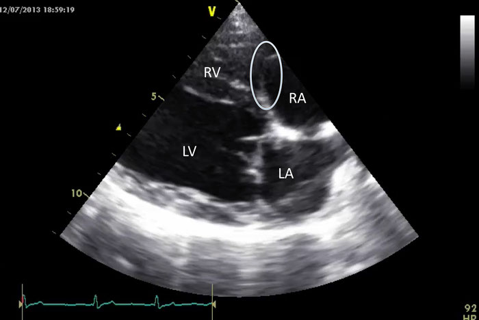
ultrasound picture of Bysterstorff's Avalon Artus (free, negativ)

ultrasound picture of Bysterstorff's Beam me up Schröder (high degree of tricuspid valve dysplasia)
Some explanations:
From this angle you see the left half of the heart from below ('lying', with left vestibule LA down right and left ventricle LV down left) and the right half of the heart at the top ('lying', with right vestibule RA up right and right ventricle RV up left).
The screenshots are labeled from us for a better understanding:
RA - right vestibule
RV - right ventricle
LA - left vestibule
LV - left ventricle
The circles mark the tricuspid valves - in case of Schröder only stumps exist, at which only one is seen at the screenshot here.
What can you see now in those two videos and the screenshot concerning the TVD?
First of all, you can see that Schröders right half of the heart (the black part on top of Schröders video and screenshot, RA+RV) is clearly bigger then Artus´ right half of the heart (the black part on top of Artus video and screenshot, RA+RV). Especially Schröders´ greatly enlarged right vestibule (RA) in comparison to his right ventricle (RV) is noticeable – in case of Artus you can see the right and healthy proportion from right vestibule (RA) to right ventricle (RV). That Schröders right vestibule is enlarged so immensely is the consequence of the blood backflow, caused by the TVD (regugitant jet, see next video).
Further, you see that the proportion of both halves of the heart (so RA+RV compared to LA+LV) greatly differs between the two dogs. As it is seen in Artus´ video and screenshot it is meant to be – right half of heart (RA+RV) as a 'small little bag' attached to the left half of heart (LA+LV), not the other way around as in the case of Schröder. This disproportion of Schröders halves of heart is even increased because the increased left part displaces the right part even further.
Illustration of the changed blood flow:
Some explanations:
Here, you can see the right half of the heart on the left side ('upright', with right vestibule RA down left and right ventricle RV up left) and the left half of the heart to the right ('upright', with left vestibule LA down right and left ventricle LV up right).
In a from cardiologist chosen image section (visible in the video) the blood is marked in terms of colors using the color Doppler ultrasound. Here the blood is either red (in direction to the transducer) or blue (going away from the transducer).
You can see the blood flow in the right half of the heart, from the right vestibule through the healthy or pathologically changed tricuspid valve to the right ventricle.
What can you detect with this video?
It is obvious that there are extreme differences in the physiological structure between Artus and Schröders tricuspid valve. While Artus´ tricuspid valve and its optimal ability to close properly is visible, Schröders tricuspid valve only exists in form of stumps and does not close at all.
Further, in case of Schröder the extreme blood backflow from the right vestibule to the right ventricle with each systole is visible in form of an extremely big blue-colored tricuspid regurgitation.